
G’day team
Moving on from our first post on the types of muscles in the body, we’ll now focus on the functional aspect of skeletal muscles. Namely, we’ll look at how muscles are arranged, their anatomy, and how they work at a cellular level, their physiology.
From here on out, every time I use the work ”muscle” I mean ”skeletal muscle”
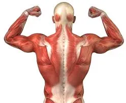
Image source
Anatomy of Muscles
Our entire skeleton is wrapped in muscles, which act not only to move our bones and allow us to mobilize but also to produce heat, to protect our organs and to stabilize our joints.
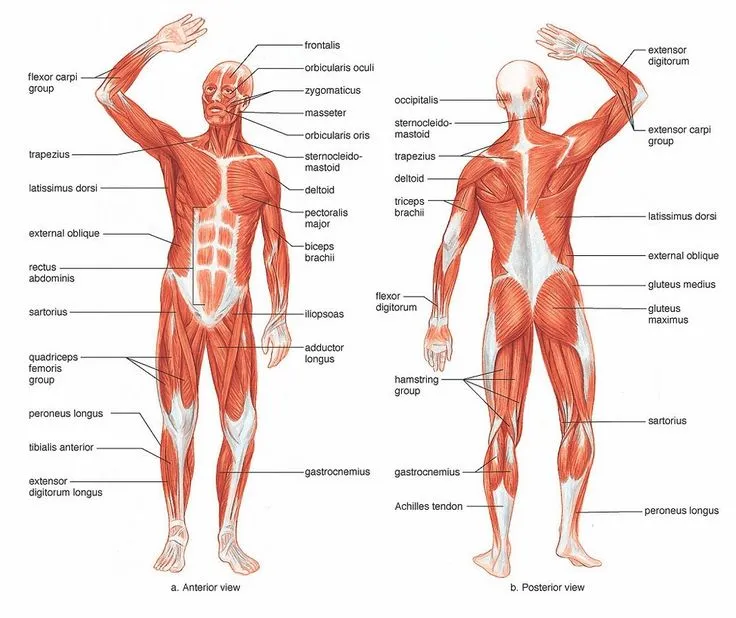
Image source
The diagram above gives a good indication of just how much muscle our body is wrapped in, but it does a poor job of indicating how many separate muscles there are. Just to give you an idea of how many muscles are used to control “fine motor skills” such as writing and typing, there are eleven muscles in the back of the forearm alone, each with a different function, and a further nine in the front compartment.
Structure of a Muscle
Muscles are best thought of as bundles, of bundles of bundles. Imaging taking a packet of spaghetti, opening it and then wrapping it in a rubber band to make bunches. Then taking 10 of these bunches and wrapping them in cord to make bundles. And taking ten of these bundles and wrapping them in rope to make… big bundle-bunch things of spaghetti. The idea is that each time we want to look at a smaller component of our new big bundle-bunch it will, in turn, be composed of even smaller components. To look at this from a muscles perspective we have…
• Actin and Myosin proteins group to make contractile units called myofibrils
• Many myofibrils are contained in muscle cells called myocytes (aka a muscle fiber)
• Many myocytes are wrapped in a thin layer of fascia to make fasciculi
• Many fasciculi are wrapped in a thick layer of fascia and tendon to make a muscle belly
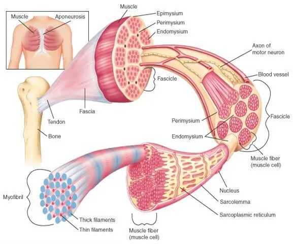
Image source
We should also take not that there are other cell types tucked away in with all those muscle fibres. Each muscle needs a blood and lymph supply so there will be arteries, veins and lymph vessels in a network through all our muscles too. Along with this our immune system has cell type called dendritic cells which have an important role in immune function and muscle repair.
How Muscles Contract
Alrighty! So now we’re all experts on the anatomy and structure of muscles, let’s have a bit of a chat about how they work.
The ability of a muscle to contract comes down to their smallest units, their actin and myosin composed myofibrils. The best way to understand a muscle contraction is to think of a centipede crawling up a ladder. With the centipede being myosin, and actin being the ladder.
Actin and myosin sit side by side in bundles, with actin anchored in place and myosin relatively free to move. Myosin is the active protein in this arrangement, covered in myosin heads which are capable of latching on to actin and then bending to drag the myosin chain along the actin.
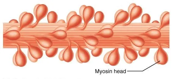
Image source
Despite the fact that myosin does all the hard work, it’s actually the actin component that controls this process. Acting fibers are dotted along their length with myosin binding sites which attract myosin heads, allowing them to latch on. But in the resting state these sites are generally covered by another long protein that wraps around actin, called tropomyosin. Just to add complexity to this mechanism, there is a third protein found at lengths along tropomyosin, called troponin, which acts as a switch or lever. When our muscles receive the signal to contract troponin pushes tropomyosin off the myosin binding sites on actin, the myosin head is then free to latch on and start its own little waltz (confused yet? :D).
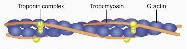
Image source
Now we get to the real nitty gritty. At the moment we’ve got a myosin head bound to its myosin binding site on an actin filament, but being stuck in this position really isn’t going to help us go anywhere.
This is where our trusty little energy packets of ATP come in! ATP is an energy carrying molecule, and it’s what allows the bending of myosin heads that pulls myosin along actin. See in its natural form each myosin head is bound to a molecule of ADP (like a de-energized form of ATP) and a single phosphate ion (known as Pi). After binding to its site on actin, the myosin head releases this ADP and Pi and uses the energy from this release to pull itself along the actin (called the ‘power stroke’). Next, a fresh new molecule of ATP binds to the myosin head and the head detaches from the myosin binding site on actin. Finally, ATP is broken (used) into ADP and Pi, re-cocking the myosin head and getting it ready for another round, it re-attaches to another myosin binding site higher up on the actin filament and the cycle starts again.
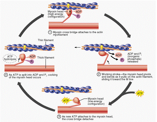
Image source
Image source
Fun Fact – Rigor Mortis
So that’s how muscles work. But I feel back drowning everyone with molecular biology terms and leaving them with nothing fun. So let’s talk about rigor mortis…
As we know, muscle contraction relies on the availability of ATP. When we die we’re no longer producing this molecule and our stores soon deplete. As our body's processes slowly break down, all the important ion channels and pumps which keep the chemical balance in our cells perfect, also breakdown. One of the things this causes is the rush of calcium in to our muscle cells. When we’re alive it’s the flooding of calcium into muscle cells that tells the cell it’s time to contract (calcium is what binds troponin, pushing tropomyosin off the myosin binding sites on actin). This calcium flood triggers the first step of contraction, and our myosin heads bind to their sites on actin and then release ADP + Pi to complete their power stroke. Unfortunately, we’ve depleted all our ATP and the myosin heads are no longer able to let go. Stuck in this ‘latched-on’ state our muscles enter a type of rigid semi-contraction that we call rigor mortis.
Rigor mortis can occur between two and six hours after death, depending on a lot of factors such as temperature, the patient’s blood glucose levels and lactic acid levels at time of death. Eventually, as part of the breaking down process of dying, the myosin heads are degraded by rogue enzymes that run rampant in cells, and bodies become flaccid once more.
Thanks
Thanks, team, I hope everyone had fun reading and learned something awesome! Be sure to stick around for part three where we'll talk about muscle growth!
Thanks
-tfc
Resources
Lubopikto
Study.com
Biology 103 - brynmawr.com
Open text
Difference between
Major Differences
Visible Body
steemstem